1
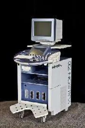
USED GE Voluson 730 EXPERT
 $
69.93
$
53.79
$
69.93
$
53.79
Title
Please select combo product attributes
The combo subtotal is $,SAVE
$
Unavailable
Quantity
-
+
ADD TO CART
BUY IT NOW
Main Features
- RealTime 4D Biopsy
- 3D Multiplanar Display
- 3D Power Doppler
- B-Flow Imaging (BF)
- CHI – Coded Harmonic Imaging
- The tissue and border differentiation are improved by the CrossBeam CRI. Added to this is a unique spatial compound acquisition and processing technique
- HD-Flow – bi-directional power Doppler allows for the vascular study to be more sensitive and cuts down the overwriting connected with the standard color Doppler
- Automatic Optimization
- VOCAL II – automated volume calculation
- RealTime 4D Biopsy
- 3D Multiplanar Display
- 3D Power Doppler
- XTD View Imaging
- Coded Contrast Imaging – Contrast Media
- Volume Contrast Imaging (VCI) improves the assessments of size, margins and internal structures of lesions through enlarged image quality in all three planes
- Speckle artifacts become decreased by the real-time software called Speckle Reduction Imaging (SRI)
- Studies for the exams and documentations become simple because of the Tomographic Ultrasound Imaging (TUI) which allows for a simultaneous view of multiple slices of volume data set
- Inversion (3D Visualization of anechoic structures)
- Coded Excitation
System Details
- Depth: 100 cm (39.4 in)
- Touch Screen: 10.4 in High Resolution color LCD screen
- Height: 142 cm (55.9 in)
- Monitor: 15” High-Resolution non-interlaced flat CRT
- Width: 68 cm (26.7 in)
- Weight (no peripherals): 300 lb (136 kg)
Operations
- Obstetrical
- Breast
- Vascular/Peripheral
- Pediatric and Neonatal
- Urological
- Abdominal
- Cardiology
- Gynecological
- Small Parts
- Transcranial
- Musculoskeletal
Transducer Types
- Microconvex Array
- Volume ‘4D’
- Sector Phased Array
- Linear Array
- Convex Array
Operating Modes
- M-Mode (M)
- Color Flow Mode (C)
- Tissue Doppler Imaging (TD)
- B-Flow (BF)
- CW Doppler (CW)
- Volume Mode (3D/4D)
- B-Mode (2D)
- M-Color-Mode (MC)
- Power Doppler Imaging (PD)
- PW Doppler with high PRF (PW)
- Extended View (XTD View)
- Coded Contrast Imaging (Contrast Media)
Transducers
- GE AC2-5 – 2-5MHz, Convex
- GE IC5-9H – 4-9MHz, Convex
- GE PA2-5P – 1-3MHz, Phased Array
- GE SP10-16 – 5-17MHz, Linear
- GE SP6-12 – 3-11MHz, Linear
- GE RAB2-5L – 1-5MHz, Convex Volume
- GE RAB4-8L – 2-8MHz, Convex Volume
- GE RIC5-9H – 3.7-9.3MHz, Convex Volume
- GE RNA5-9 – 3-9MHz, Convex Volume
- GE M7C-H – 3-8MHz, Convex Matrix Array (BT05 and up)
- GE AB2-7 – 2-7MHz, Convex
- GE IC5-9 – 3.3-10.0MHz, Convex
- GE 4C-A – 1-5MHz, Convex (BT05 and up)
- GE PA6-8 – 4-10MHz, Phased Array
- GE SP4-10 – 3-8MHz, Linear
- GE RAB2-5 – 2-5MHz, Convex Volume
- GE RAB4-8P – 2-7MHz, Convex Volume
- GE M12L-H – 5-13MHz, Linear Matrix Array (BT05 and up)
- GE RIC5-9 – 3.3-10.0MHz, Convex Volume
- GE RRE6-10 – 4-9MHz, Convex Volume
- GE RSP5-12 – 3.5-11.0MHz, Linear Volume
You may also like